|
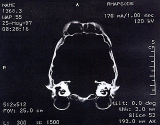
USE OF CT-IMAGING & 3-D COLOR RECONSTRUCTION FOR COMPARISON OF CRANIAL DENSITY IN CAPTIVE & WILD CHEETAHS (Acinonyn jubatus) by D.A.Fagan, D.D.S., J.E.Oosterhuis, D.V.M., C.V.Fiorello, MS., G.F.Daleo, & J.E.Fagan Memo to the file : For several years it has been apparent that the F.P.E. lesions in the cheetah are related to an "arch length discrepancy" problem – that is a dis-harmony between the length of , or mass of alveolar bone available to support the individuals dentition, . . . and . . . the total mass of dentition or total length of all of the teeth. The question has remained " how to identify the cause of the problem "? Clearly, it was necessary to look at, evaluate and compare a variety of cheetah skulls. This is more easily said than done, because of the C.I.T.E.S. restrictions on the importation of endangered species or their "body parts". It has therefore taken a number of years to manage to gather a representative sample of cheetah cranial material together, and to conduct a reproducible analysis. The material presented here represents the results of such an analysis. This material was presented at the 1997 meeting of the American Association of Zoo Veterinarians - poster session. The material is presently under analysis and will soon be assembled in the form of a formal paper for publication. The color frames boarding the various sections are the 3-D color reconstructions of the individual CT-Scan radiographs. The "white" portion of each image represents the x-ray portion of that numbered "slice". Figures 1 and 2 are but two of the original CT-Scan radiographs from which the 3D images were reconstructed.
Figure 1
Figure 2
|
|
|
For translation information and instructions, please CLICK HERE. http://www.colyerinstitute.org
This web site is best viewed with a browser setting of 800 x 600 or greater. If you use a smaller screen size and would like to view the content of this site using an alternate interface, please click here. |